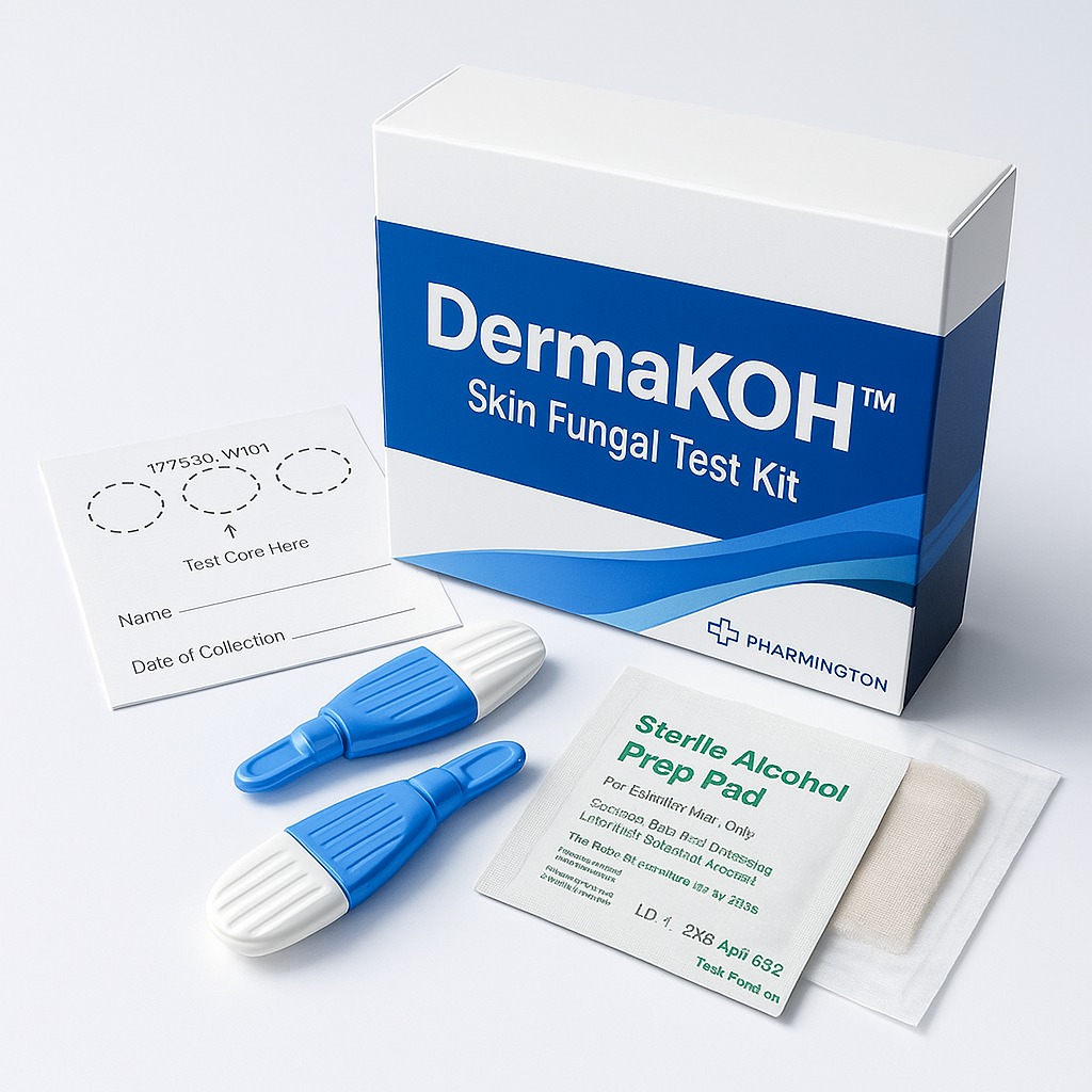
DermaKOH™ is a ready-to-use diagnostic kit designed for rapid detection of dermatophyte, yeast, and fungal infections in skin, hair, and nails. Developed for dermatologists, clinics, and advanced skin labs, it provides high accuracy, clarity, and convenience in one compact sterile package.
Each kit includes pre-measured 10% KOH reagent, sterile glass slides, and applicators for immediate use — ensuring reliable results and safe handling.
Key Features & Benefits:
Accurate identification of fungal elements (hyphae, spores, yeast cells)
Ready-to-use 10% KOH solution with dropper precision
High-clarity glass slides for microscopy
Leak-proof, single-use reagent vials
Compact, hygienic, and easy to store
Kit Contents:
5 × 1 mL KOH reagent vials (10%)
5 × Sterile glass slides
5 × Wooden applicators
Instruction leaflet with illustrated procedure
Biohazard disposal pouch
Usage:
Apply specimen to slide → add 1–2 drops of KOH → cover and observe under microscope after 5–10 minutes.
£61.50
Fungal infections of the skin, hair, and nails represent some of the most prevalent dermatologic conditions encountered in clinical practice worldwide. Dermatophytes, yeasts, and non-dermatophyte molds are responsible for a wide spectrum of superficial mycoses, often presenting with overlapping clinical features that complicate diagnosis based solely on visual inspection. (1)
Despite advances in dermatologic imaging and molecular diagnostics, potassium hydroxide (KOH) microscopy remains a cornerstone of fungal detection due to its simplicity, cost-effectiveness, and rapid turnaround time. However, diagnostic accuracy is highly dependent on preparation quality, reagent concentration, and procedural consistency. (2)
DermaKOH™ Skin Diagnostic Kit has been developed to address these variables by providing a standardized, ready-to-use diagnostic solution designed specifically for dermatologists, clinics, and advanced skin laboratories. This review examines the scientific rationale, diagnostic methodology, and clinical relevance of DermaKOH™ within modern dermatologic practice.
The integumentary system provides a nutrient-rich environment for fungal colonization, particularly within keratinized tissues. Dermatophytes exhibit a unique affinity for keratin, thriving within the stratum corneum, hair shafts, and nail plates. (3)
Keratin acts as both a physical barrier and a metabolic substrate, shielding fungal elements from immune surveillance while limiting visibility during routine examination. Consequently, effective diagnostic techniques must selectively dissolve keratin structures without compromising fungal morphology. (4)
This fundamental biological challenge underpins the continued relevance of KOH-based diagnostic methods in dermatology.
Potassium hydroxide functions by digesting keratin and cellular debris through alkaline hydrolysis, rendering fungal structures such as hyphae, pseudohyphae, spores, and yeast cells optically visible under light microscopy. (5)
Importantly, fungal cell walls—composed primarily of chitin and glucans—remain intact under KOH exposure, allowing for clear differentiation between host tissue and pathogenic organisms. (6)
KOH preparation has been recognized for decades as a diagnostic gold standard in the identification of superficial mycoses. Multiple clinical guidelines continue to recommend KOH microscopy as a first-line diagnostic approach in suspected fungal infections due to its speed and diagnostic reliability when properly performed. (7)
While traditional KOH microscopy is effective, variability in reagent concentration, handling techniques, and slide quality can significantly affect diagnostic outcomes. DermaKOH™ has been engineered to minimize these limitations through a standardized, ready-to-use design.
DermaKOH™ integrates:
This formulation-centric approach reflects modern diagnostic principles emphasizing reproducibility, safety, and operator convenience. (8)
The use of a standardized 10% KOH solution represents a clinically validated balance between efficacy and tissue preservation. Concentrations within this range have been shown to effectively clear keratinized material while maintaining the structural integrity of fungal elements. (9)
Excessively concentrated solutions may distort morphology, whereas lower concentrations may lead to incomplete keratin digestion and reduced sensitivity. The DermaKOH™ formulation aligns with established dermatologic best practices. (10)
DermaKOH™ is designed for the rapid evaluation of suspected fungal infections affecting:
Its applications extend across multiple clinical environments, including dermatology clinics, aesthetic practices, and advanced skin diagnostic laboratories. The kit supports immediate point-of-care decision-making without reliance on extended culture-based methods. (11)
One of the primary strengths of DermaKOH™ lies in its ability to enhance diagnostic clarity. By standardizing reagent quality and slide preparation, the kit reduces procedural variability, a known source of false-negative results in routine microscopy. (12)
Clear visualization of hyphae, spores, and yeast cells improves diagnostic confidence and supports appropriate therapeutic decision-making.
DermaKOH™ is intended исключительно for external diagnostic use. Safety is supported by:
When used according to instructions, KOH-based diagnostics demonstrate a favorable safety profile with minimal risk to clinicians and patients. (13)
DermaKOH™ should be viewed as a diagnostic aid that complements, rather than replaces, advanced diagnostic modalities such as fungal culture, histopathology, or molecular testing. Its rapid results make it particularly valuable in initial assessments and treatment planning. (14)
Within evidence-based dermatology, standardized point-of-care diagnostics play an increasingly important role in improving efficiency, accuracy, and patient outcomes.
DermaKOH™ Skin Diagnostic Kit exemplifies a modern, standardized approach to KOH-based fungal diagnostics. By combining a clinically validated 10% KOH formulation with sterile, ready-to-use components, DermaKOH™ enhances diagnostic accuracy, procedural consistency, and clinical convenience.
Grounded in established dermatologic science, DermaKOH™ supports timely and reliable identification of fungal infections, reinforcing its value as an essential diagnostic tool in contemporary skin care practice.
What is DermaKOH™ used for?
DermaKOH™ is used for the rapid microscopic detection of fungal elements in skin, hair, and nail samples.
Is DermaKOH™ a treatment?
No. DermaKOH™ is a diagnostic kit and does not treat fungal infections.
Who should use DermaKOH™?
It is intended for dermatologists, trained healthcare professionals, and clinical laboratory personnel.
Does DermaKOH™ replace fungal culture?
No. It serves as a rapid first-line diagnostic tool and may be used alongside confirmatory tests when needed.
1. Hay RJ, Ashbee HR. Fungal infections. British Journal of Dermatology, 2017.
DOI: 10.1111/bjd.15842
2. Gupta AK, Foley KA. Antifungal treatment and diagnostic challenges. Dermatologic Clinics, 2015.
DOI: 10.1016/j.det.2014.09.004
3. Weitzman I, Summerbell RC. The dermatophytes. Clinical Microbiology Reviews, 1995.
DOI: 10.1128/CMR.8.2.240
4. Elewski BE. Onychomycosis: Pathogenesis and diagnosis. Journal of the American Academy of Dermatology, 1998.
DOI: 10.1016/S0190-9622(98)70447-X
5. Larone DH. Medically Important Fungi: A Guide to Identification. ASM Press, 2011.
6. Gow NAR, Latge JP, Munro CA. The fungal cell wall. Microbiology Spectrum, 2017.
DOI: 10.1128/microbiolspec.FUNK-0035-2016
7. Gupta AK, Versteeg SG. Dermatophytosis diagnostics. Journal of Cutaneous Medicine and Surgery, 2017.
DOI: 10.1177/1203475417727636
8. Nenoff P, et al. Methods for laboratory diagnosis of dermatomycoses. Mycoses, 2014.
DOI: 10.1111/myc.12223
9. Roberts DT, Taylor WD, Boyle J. Guidelines for diagnosis of superficial fungal infections. British Journal of Dermatology, 2003.
DOI: 10.1046/j.1365-2133.2003.05678.x
10. Hainer BL. Dermatophyte infections. American Family Physician, 2003.
PMID: 12643357
11. Drake LA, et al. Guidelines of care for superficial mycotic infections. JAAD, 1996.
DOI: 10.1016/S0190-9622(96)90773-6
12. Summerbell RC. False-negative results in KOH microscopy. Medical Mycology, 1997.
DOI: 10.1080/02681219780000781
13. CDC. Guidelines for safe laboratory handling of chemicals. Centers for Disease Control and Prevention.
14. Nenoff P, Krüger C. Modern diagnostics in dermatology. Dermatology Practical & Conceptual, 2014.
DOI: 10.5826/dpc.0402a03
© 2025 Pharmington. All rights reserved.